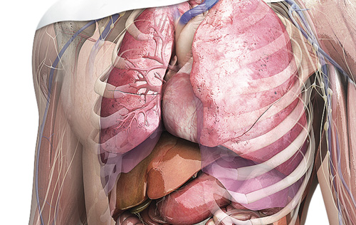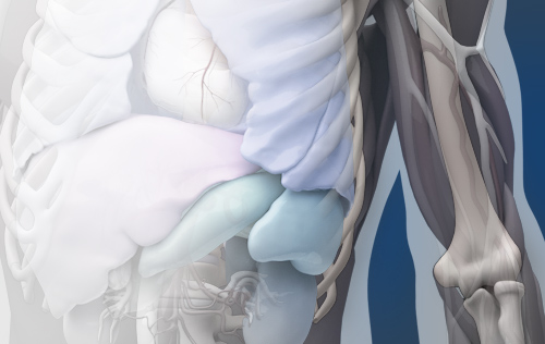
Solid 3D Male Circulatory System
Need to model a system involving the Blood Flow or the Heart?
Zygote's detailed Solid 3D Male Circulatory System is perfect for:
- Catheter or Stent Engineering
- Cardiac Fluid Dynamics Studies
- Simulating Cardiac Interventions & Treatments
- CAD Product & Device Design
Developed from medical scans, these high fidelity 3D geometries of the Circulatory System can be depended on for accurate results.
Includes the Zygote Solid 3D Heart Model.
A free eDrawing of this model is available. What is an eDrawing? Click here to find out more.
Zygote's Solid 3D Anatomy has set the standard in CAD and Simulation for over a decade.
- Formats:
- ProE/Creo
- IGES
- ParaSolid
- SolidWorks
- Step
- Delivery Method: Download
- Price: $4,800
"I want to thank . . . the team at Zygote for proving us with the most complete and easy to use human body data-set on the market today. I was extremely impressed with the accuracy of the geometry and the detail of the textures, which gave Rushes a solid base on which to design the visual effects for "Human Body:Pushing The Limits" The continued support and upgrading of the model library, allowed us to push the creativity of our shots and surpass the expectations of our client."
Hayden Jones / VFX Supervisor - Rushes Postproduction Ltd.
The Solid 3D Male Circulatory System includes the heart and pericardium as well as blood vessels serving the heart, head, trunk, pelvis, arms and legs. Blood vessel interior diameters were acquired through use of scans leveraging contrasting agent. Diameters, contours and wall thickness of the blood vessels of the heart, neck, vena cava, aorta and proximal femoral arteries conform to data obtained from medical scans. Vessel wall thickness throughout the rest of the body was developed by projecting the same ratio of interior vessel diameter to blood vessel wall thickness as observed in scans, down through the extremities.
The heart is a component of a larger anatomy collection that includes anatomical geometries taken from different scan subjects and integrated into a whole-body assembly of a 50th percentile (U.S. by height and weight) Caucasian male. Full-body scan data of this individual provided the over-all template or infrastructure for the entire assembly of the circulatory system constructed from various scan inputs - including contrast-dye scans of different scan subjects. The Heart was developed from an excellent heart scan of a subject different than the primary template subject. The resultant heart data was subsequently positioned, without changing the proportions or shape, to fit the 50th percentile male scan data.


