Solid 3D Male Organs
Simulating a surgical procedure or the impact a system will have on the organs of the body?
You need Zygote's Solid 3D Male Organs.
Zygote's Solid 3D Male Organs are perfect for:
- Surgical Simulation
- Medical Device Design
- Injury & Trauma Modeling
- Whole-Body Simulation
Use Zygote's highly detailed organs or simplified stand-in shapes to get results you can rely on.
A free eDrawing of this model is available. What is an eDrawing? Click here to find out more.
Zygote's Solid 3D Anatomy has set the standard in CAD and Simulation for over a decade.
- Formats:
- ProE/Creo
- IGES
- ParaSolid
- SolidWorks
- Step
- Delivery Method: Download
- Price: $5,040
"We wanted to demonstrate the kind of 3D experience we should all expect from modern browsers. We chose the human body because of how fascinating it is to learn about it, and we chose to work with Zygote Media Group because of their outstanding imagery and team."
Roni Zeiger, MD / Google's Chief Health Strategist
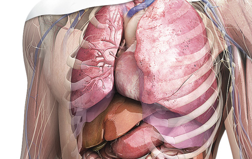
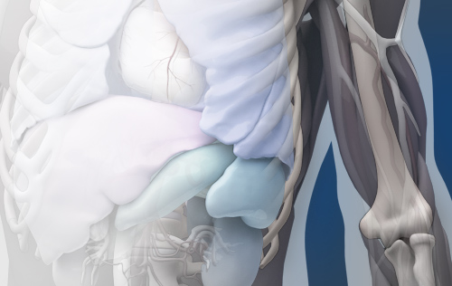
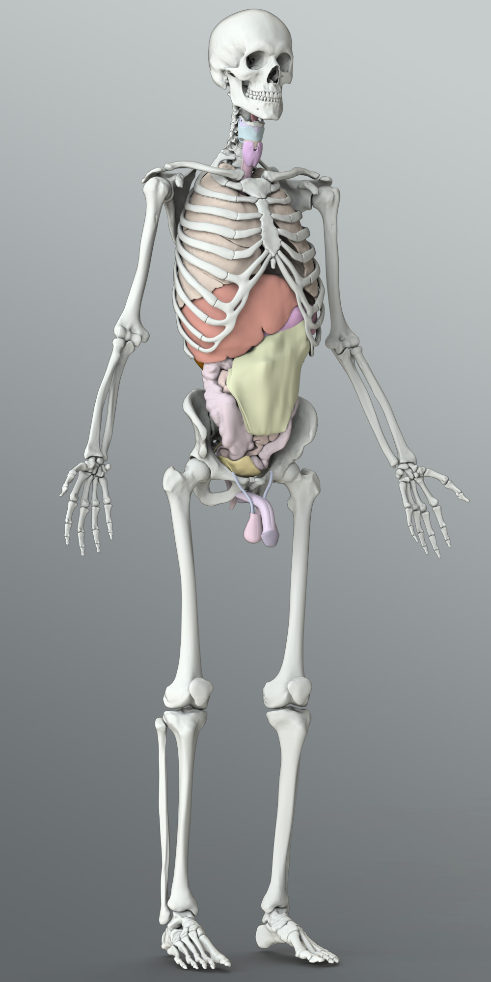
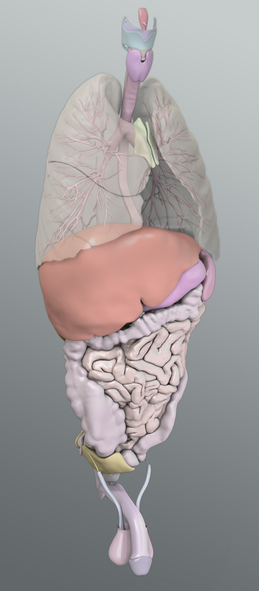
The Zygote Solid 3D Male Organs were developed using a CT scan of a 50th percentile male individual as source material. Interior cavities exist for the Trachea and Bronchioles, the Esophagus, Stomach and Intestines, the Renal pelvis, the Urinary bladder and the Gall bladder. The Greater omentum is a simplified geometry created by hand using atlases as reference, to provide appropriate mass and structure to the abdomen and digestive organs located therein. Though a thoracic organ, the Solid 3D Heart is not included in this collection.
Organs of the Respiratory System
The Organs and features included from the Respiratory System include detailed Tracheal and Bronchial anatomy which correspond brilliantly with the branching vessels of the Pulmonary Artery and Veins (part of the Solid 3D Male Circulatory collection and not included in this collection). The detail and fidelity of the lung tissue is very high and correlates well with the Skeleton and Muscle System (licensed separately) – with Costal grooves fitting the ribs. Pleura fitting the lungs are also available as a light standing for whole-body modeling.
Digestive System Organs
The Digestive System's organs, including the Esophagus, Stomach, Small and Large Intestines, as well as the other abdominal organs were created using scan data of the initial scan specimen and digitized by medical modelers. The interior digestive cavity is detailed in the stomach, clearly showing rugae, while the interiors of the large and small intestines are created procedurally by extrusion of the outer surface to create the lumen of the intestinal cavities with appropriate wall thicknesses as observed from scan data.
Organs of the Endocrine, Urinary and Reproductive Systems
The organs or glands of the Endocrine system that are included in this assembly are: the Thyroid, Thymus and Adrenal glands and the Pancreas; each constructed using scan data as primary source. The Pituitary and Pineal glands are not included in this collection, but they do exist in the Nervous System collection. Testicles are included in this collections, however, the geometry was constructed by medical modelers using visual reference from atlases and publications as reference.
High fidelity geometry defines the exterior and interior of the Urinary bladder, Kidney exterior, Collecting ducts and Renal pelvis and Ureters.
The organs of the Male Reproductive System were developed from visual reference include the Penis, Scrotum, Testis, Epididymis, Prostate, Seminal vesicle and Ductus deferens.
Each structure and component is an individual object organized in the assembly for quick access and use.


