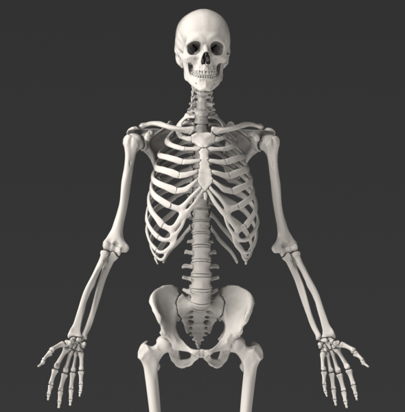
Solid 3D Male Skeleton
One of our most popular collections due to the broad range of applications, this Skeletal System will impress you.
Zygote's Solid 3D Male Skeleton is perfect for:
- Kinematic Studies
- Orthopedic Device Design
- Stress & Force Research
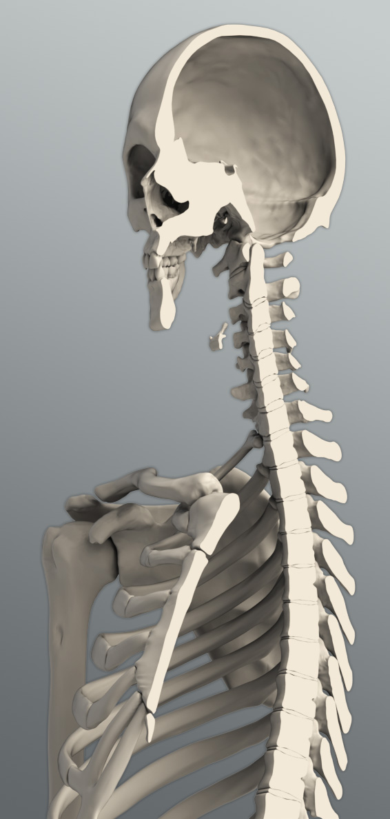
The Zygote Solid 3D Skeleton Model sets a new benchmark for solid human anatomy. It is perfect for orthopedic design, animation, marketing renders, or finite element analysis. It was developed from CT scans of a 50th percentile male, and was carefully modeled to retain subtle anatomical nuances unique to specific bones. The skeleton is a solid assembly of individual bones, intervertebral discs, and teeth, making selection and manipulation of individual components simple and straightforward.
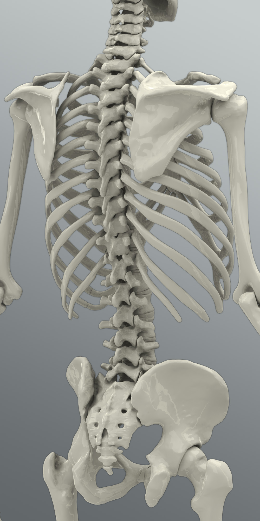
The Zygote Solid Skeleton model has a high level of geometric detail while efficiently maintaining anatomical fidelity throughout the model. An example of the great improvements in this version of the solid skeleton is the skull; which includes all major foramina, an accurate interior to the cranium, and each sinus cavity.
Solid Skeleton Component Products
- Solid Skeleton Spine Model - $ 3,350
- Solid Skeleton Skull Model - $ 1,900
- Solid Skeleton Limbs - $ 1,900
A free eDrawing of this model is available. What is an eDrawing? Click here to find out more.
Zygote's Solid 3D Anatomy has set the standard in CAD and Simulation for over a decade.
- Formats:
- ProE/Creo
- IGES
- ParaSolid
- SolidWorks
- Step
- Delivery Method: Download
- Price: $6,090
"The Zygote model was perfect for all of our needs, and really came through for us in crunch time. The model itself was beautifully detailed and thorough. The separate systems were set up exactly as we needed them so that we could turn certain systems off and on with ease. The builds were so organized, we had no problems retexturing and isolating specific parts of the model. We were all very pleased and very impressed with the build."
Brad Powell / VFX Supervisor - CSI:NY


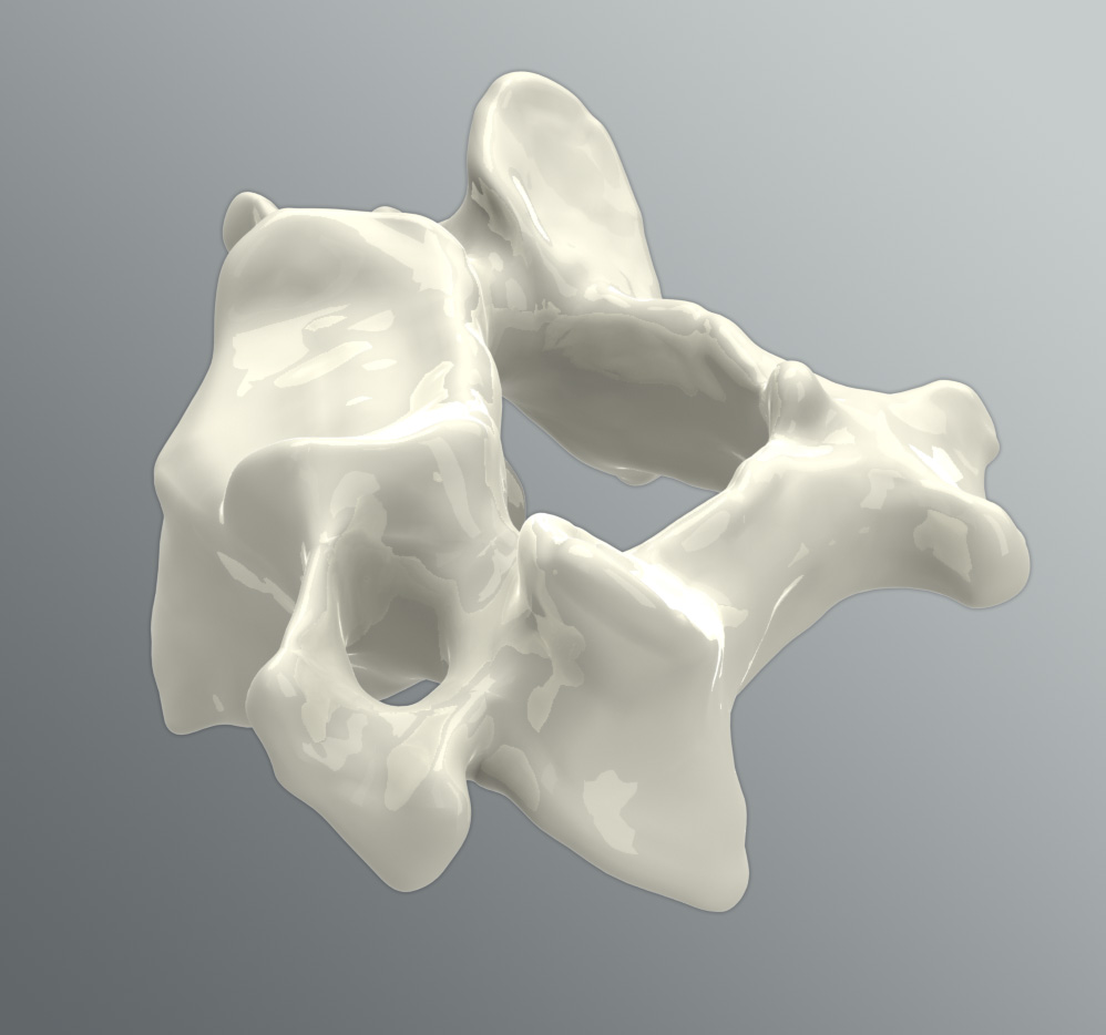
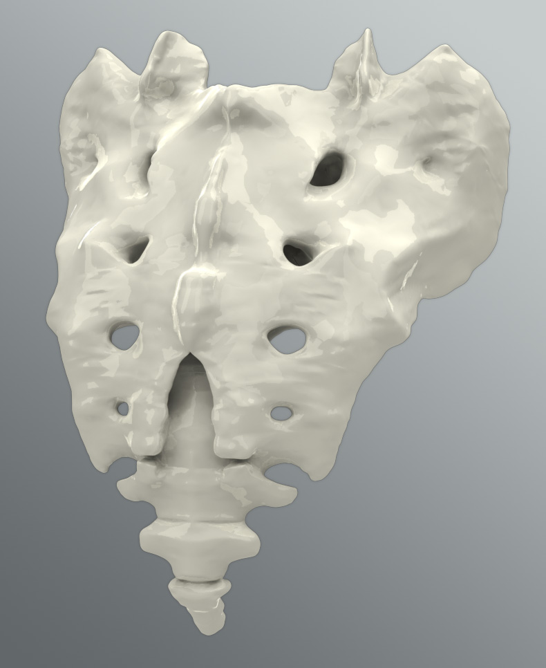
The skeleton is created entirely from CT scans of one 50th percentile individual as detailed below. High fidelity geometries detail major features of each bone including fossae, foramina, tubercles and tuberosities. Each bone is a separate part, labeled with conventional nomenclature. The skeleton is symmetrical with the scan subject's data being mirrored about the central axis. Cortical and cancellous bone tissue layers are licensed separately and available for select bones upon request.
Since the scan posture was recumbent, adjustments were made to alter the posture of the skeleton to standing. Careful attention was paid to preserve bone shape, articulations and the spatial relationships between bones. Standing Cyber scan data of the CT scan subject were taken for correct standing posture and external landmarks were used to adjust the posture from recumbent posture to standing.
Included in this collection are the intervertebral discs.
Creators of the world's leading 3D human anatomy models for use in medical illustration, animation, engineering, simulation, and anatomy software products.
2015 © All Rights Reserved. Privacy Policy | Terms of Service | Careers