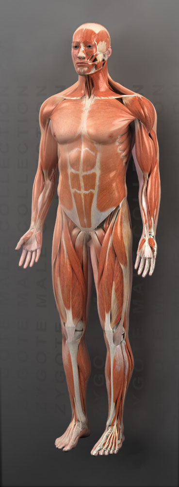
3D Male Muscular System
- Formats:
- 3D Studio Max
- Blender (OBJ)
- Cinema 4D
- Generic OBJ
- Maya
- Polygons (as tris): 609610
- UV Coordinates: Available
- Textures: Available
- Grouping: Yes
- Delivery Method: Download
- Price: $6,560
- Rigged: No
+1 (801) 765-4141
or
3D Male Muscular System
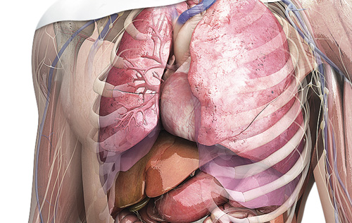
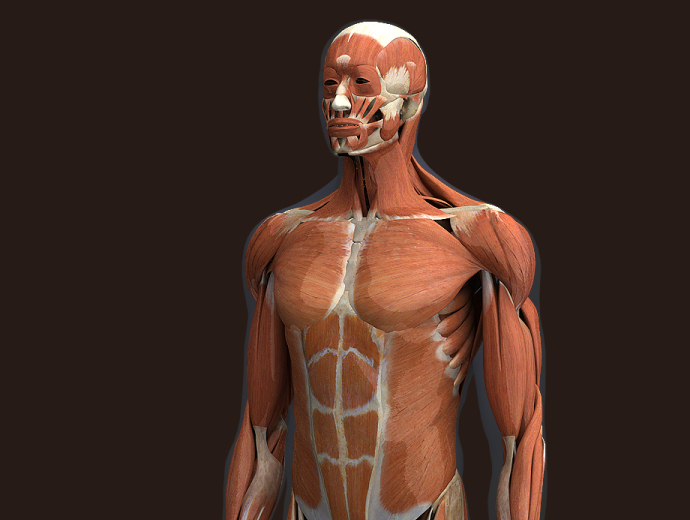
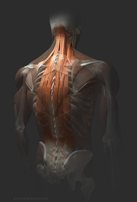
The Zygote 3D Male Muscular System Model is one of our flagship products. It includes each individual muscle of the axial and appendicular skeleton, including deep muscles of the back and spine with proper origin and insertion points. Animators using this product can easily create animations and images showing any variety of muscle configurations such as: entire muscle system, agonist and antagonist muscle combinations or each muscle flexing individually.
The 3D Male Muscle System is a superior product created through years of development working with scanned human forms, photographic and illustrative anatomical atlases. Texture maps were derived from photographs of human cadaver muscle tissues, enabling the user to create photo-real images and animations. The system includes all major muscles of the axial and appendicular skeleton, both superficial and deep. The 3D Muscle System has been geometrically optimized to minimize render times while maximizing shape and surface integrity. Each muscle is volumetric and individually grouped for quick selection.
Features of the 3D Muscle System include:
- Accurate, space-filling/volumetric treatment of each muscle
- Each individual muscle grouped for selection and visualization
- Accurate origin and insertion points for each muscle
- Compatible with Zygote 3D Male Anatomy Collection
- Compatible with other Zygote 3D Male Anatomy Skeletal and Other Systems
- Optional Texture maps for the 3D Male Muscular System model are available. The various regions of the muscular system are divided up into twenty 2048 x 2048 color maps.
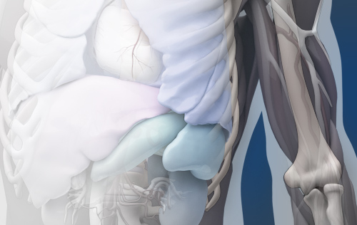
The skeletal system is tightly integrated with muscular system as shown in many of the above images. These two systems complement each other very well. The skeletal system is sold separately.
Head A
- left and right auricularis anterior
- left and right auricularis posterior
- left and right auricularis superior
- left and right buccinator
- left and right corrugator supercilii
- left and right deep masseter
- left and right superficial masseter
- left and right depressor anguli oris
- left and right depressor labii inferioris
- left and right depressor supercilii
- left and right digastric
- left and right fibrous loop for intermediate digastric tendon
- left and right hyoglossus
- left and right levator anguli oris
- left and right levator labii superioris
- left and right levator labii superioris alaeque nasi
- left and right orbicularis oculi
- left and right risorius
- left and right stylohyoid
- left and right temporalis
- left and right thyrohyoid
- left and right zygomaticus major
- left and right zygomaticus minor
- left and right internal pterygoideus
- left and right external pterygoideus
Head B
- left and right alar nasalis
- left and right transverse nasalis
- depressor septi nasi
- frontalis
- mentalis
- mylohyoid
- nasal cartilage
- orbicularis oris
- procerus
- left and right styloglossus
- left and right geniohyoid
- left and right hyoglossus
Neck A
- left and right levator scapulae
- left and right scalene anterior
- left and right scalene middle
- left and right scalene posterior
- left and right semispinalis capitis
- left and right semispinalis cervicis
Neck B
- left and right omohyoid
- left and right platysma
- left and right splenius capitis
- left and right sternocleidomastoid
- left and right sternohyoid
- left and right sternothyroid
- left and right subclavian
- genioglossus
- left and right styloglossus
- left and right hyoglossus
- left and right geniohyoid
- mylohyoid
TorsoMid
- external abdominal oblique
- rectus abdominus
- transverses abdominus
- trapezius
Torso A
- left and right coracobrachialis
- left and right infraspinatus
- left and right latissimus dorsi
- left and right pectoralis major
- left and right pectoralis minor
- left and right serratus anterior
- left and right subscapularis
- left and right supraspinatus
- left and right teres major
- left and right teres minor
Torso B
- left and right iliocostalis
- left and right intercostal
- left and right internal abdominal oblique
- left and right longissimus
- left and right quadratus lumborum
- left and right spinalis
- left and right rhomboid major
- left and right rhomboid minor
Diaphragm
Back
- left and right longissimus thoracis
- left and right spinalis thoracis
- left and right iliocostalis
- left and right spinalis cervicis
- left and right longus colli
- left and right multifidi
Back 2
- Serratus posterior inferior
- Serratus posterior superior
Arm A
- left and right abductor digiti minimi (finger)
- left and right abductor pollicis brevis
- left and right abductor pollicis longus
- left and right adductor pollicis
- left and right anconeus
- left and right bicipital aponeurosis
- left and right dorsal interosseous muscles (1,2a,2b,3a,3b,4a,4b)
- left and right extensor pollicis brevis
- left and right flexor digiti minimi brevis (finger)
- left and right flexor pollicis brevis
- left and right flexor pollicis longus
- left and right flexor retinaculum
- left and right interosseous membrane
- left and right lumbrical muscles (1,2,3a,3b,4a,4b)
- left and right opponens digiti minimi left and right opponens pollicis
- left and right palmar interosseous muscles (1,2,3)
- left and right palmaris brevis
- left and right pronator quadratus
- left and right pronator teres
- left and right supinator
Arm B
- left and right extensor digiti minimi
- left and right extensor digitorum
- left and right extensor indicis
- left and right extensor pollicis longus
- left and right flexor digitorum profundus
- left and right flexor digitorum superficialis
- l eft and right palmaris longus
Arm C
- left and right brachioradialis
- left and right extensor carpi radialis brevis
- left and right extensor carpi radialis longus
- left and right extensor carpi ulnaris
- left and right flexor carpi radialis
- left and right flexor carpi ulnaris
Arm D
- left and right deltoid
- left and right lateral biceps brachii
- left and right medial biceps brachii
Arm E
- left and right brachialis
- left and right triceps brachii lateral
- left and right triceps brachii longus
- left and right triceps brachii medius
- left and right triceps brachii tendon
Hip
- left and right gluteus maximus
- left and right gluteus medius
- left and right gluteus minimus
- left and right iliacus
- left and right inferior gemellus
- left and right obturator externus
- left and right obturator internus
- left and right piriformis
- left and right psoas major
- left and right psoas minor
- left and right quadratus femoris
- left and right sacrotuberous ligament
- left and right superior gemellus
Pelvic Floor
- Levator ani – pubococcygeus part
- Levator ani – iliococcygeus part
- Coccygeus
Leg A
- left and right adductor longus
- left and right adductor magnus
- left and right biceps femoris short head
- left and right plantaris
- left and right popliteus
- left and right tensor fasciae latae
- left and right vastus intermedius
- left and right vastus medialis
Leg B
- left and right adductor brevis
- left and right biceps femoris long head
- left and right gracilis
- left and right pectineus
- left and right rectus femoris
- left and right sartorius
- left and right semimembranosus
- left and right semitendinosus
- left and right vastus lateralis
Low Leg
- left and right abductor digiti minimi (toe)
- left and right abductor hallucis
- left and right adductor hallucis
- left and right Achilles tendon
- left and right extensor digitorum brevis
- left and right extensor digitorum longus
- left and right extensor hallucis brevis
- left and right extensor hallucis longus
- left and right flexor digiti minimi brevis
- left and right flexor digitorum brevis
- left and right flexor digitorum longus
- left and right flexor hallucis
- left and right flexor hallucis brevis
- left and right gastrocnemius
- left and right interosseous dorsal
- left and right interosseous membrane
- left and right interosseous plantar
- left and right lumbrical
- left and right patellar ligament
- left and right peroneus brevis
- left and right peroneus longus
- left and right peroneus tertius
- left and right quadratus plantae
- left and right soleus
- left and right tibialis anterior
- left and right tibialis posterior
The Zygote model was perfect for all of our needs, and really came through for us in crunch time. The model itself was beautifully detailed and thorough. The separate systems were set up exactly as we needed them so that we could turn certain systems off and on with ease. The builds were so organized, we had no problems retexturing and isolating specific parts of the model. We were all very pleased and very impressed with the build.

