
3D Kidney Cross Section
The kidney cross section 3D model includes highly detailed representations of the major structures of human kidney. Each feature is individually grouped for easy access, and a high-quality texture map is included with the model to illustrate the cut-surface anatomy of the kidney's interior.
Zygote's Solid 3D Anatomy has set the standard in CAD and Simulation for over a decade.
- Formats:
- 3D Studio Max
- Blender (OBJ)
- Cinema 4D
- Generic OBJ
- Maya
- Polygons (as tris): 34639
- UV Coordinates: Yes
- Textures: Yes
- Grouping: Yes
- Delivery Method: Download
- Price: $555
- Rigged: No
"I want to thank . . . the team at Zygote for proving us with the most complete and easy to use human body data-set on the market today. I was extremely impressed with the accuracy of the geometry and the detail of the textures, which gave Rushes a solid base on which to design the visual effects for "Human Body:Pushing The Limits" The continued support and upgrading of the model library, allowed us to push the creativity of our shots and surpass the expectations of our client."
Hayden Jones / VFX Supervisor - Rushes Postproduction Ltd.


The anatomy included in this kidney cross section model begins with hilum, or opening of the medial surface of the kidney, which serves as a portal for the ureter, the renal artery and the renal vein. This model includes a sectioned view of the ureter at the renal pelvis to provide a view inside the structure. The surface of the sectioned kidney face properly illustrates the renal cortex, renal medulla (made up of striated renal pyramids), and their relation to the circulation within the kidney. This model is a must for anyone interested in teaching or learning the basics of the Urinary System.
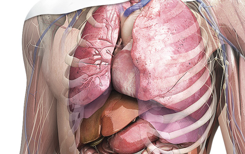
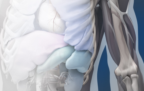
Creators of the world's leading 3D human anatomy models for use in medical illustration, animation, engineering, simulation, and anatomy software products.
2015 © All Rights Reserved. Privacy Policy | Terms of Service | Careers